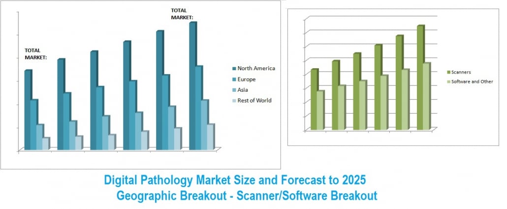
Digital Pathology Market 2020-2025
SKU 20-52
Description
Digital pathology is the technique of analyzing high-resolution digitally scanned histology images, which may then be analyzed using computational tools and algorithms. Digital pathology is enabled in part by virtual microscopy, a method of capturing microscopic images and transmitting them over computer networks. This allows independent viewing of images by large numbers of people in diverse locations.

Digital camera equipped microscopes may zoom onto a specific region on a slide to provide higher resolution of the area. In another strategy, the whole histology glass slide is scanned with the help of a high-resolution image scanner. In both cases, a pathologist reviews the digital image information to arrive at a test result. The role of computer-based imaging is very important and helps pathologists in making a diagnostic decision. Therefore, digital pathology is an intersection of traditional pathology and computers that is capable of replacing the conventional microscope-based diagnosis in the near future. Digital imaging combined with the use of powerful bioinformatics tools promise to radically change the future of pathology.

Kalorama has covered digital pathology markets in line with its coverage of related in vitro diagnostic tests for over a decade. This 2020 report clarifies the market situation given recent changes due to COVID-19.
The report provides:
- Market for Digital Pathology, Now and in 2025
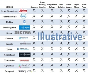
- Position of Major Players
- Regional Breakout
- Trends – Big Data
- Trends – COVID-19 and Social Distancing Impact
- Trends – AI
- Scanners Market
- Software and Other Supplies Market
- Competitors in the Market and Recent Moves: Roche, Philips, Leica, Danaher, Sectra and Others
Advances in slide scanning; digital photography and the decreasing cost of digital cameras are revolutionizing traditional ways of documenting pathology findings, collaborating on slide review, and analyzing images. There is considerable skill involved in the processing and staining tissues and fluids for investigation. Digital histology allows pathologists to collaborate in reviewing slides or send images to experts for a first reading or second opinion. This report provides insights into relevant trends to assess business opportunities in the future.
-
Executive Summary
- Introduction
- Size and Growth of the Market
- COVID-19 and Digital Pathology
-
Digital Pathology Trends
- Digital Pathology: End User Point of View
-
Data storage affordability
- Regulatory Considerations Drive Storage Purchases
- After the scan – The “Big Data” Vision of Digital Pathology
- Communications Technology As an Enabler
-
Competitive and Market Analysis
- Roche
-
Philips
- Visopharm Partnership
- Philips-Dako
-
Agilent
- ASI Agreement
- Visiopharm
-
Leica BioSystems (Danaher)
- Perio AT2 DX
-
Market Outlook
- DPA Academy Project
- Size and Growth of Market
